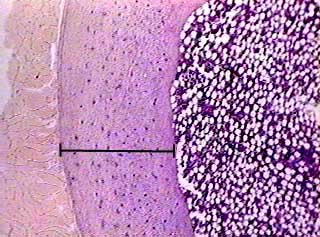
Cross Section Of A Compact Bone : Cartilage Bone Ossification The Histology Guide : To know the architecture of compact and spongy (cancellous) bone.. Bones that are longer than they are wide are called long bones. (micrograph provided by the regents of university of michigan. This is a cross section through decalcified bone. Compact bone, dense bone in which the bony matrix is solidly filled with organic ground substance and inorganic salts, leaving only tiny spaces that contain the osteocytes, or bone cells. From wikimedia commons, the free media repository.
As the names suggest compact bone looks compact and the spongy bone looks like sponges. The innermost layer of membrane is made up of. Jump to navigation jump to search. Bones that are longer than they are wide are called long bones. Compact bone is organized into parallel columns, known as haversian systems, which run lengthwise down the axis of long bones.
Bone Compact Decalcified C S from www.austincc.edu As the names suggest compact bone looks compact and the spongy bone looks like sponges. Haversian systems comprise concentric rings of bone around a central channel or haversian canal. There are two ways to study bone histology. Compact bone makes up the dense outer layer of bones. Cross section of compact bone. Osteocyte processes lie in tiny canals (canaliculi) in the bone matrix. In the center of each osteon is the central canal, a space that houses blood vessels and nerves that supply bone. In three dimensions an osteon is cylindrical in shape.
Compact bones make up 80 percent of the human skeleton;
Cross section of compact bone. As seen in the image below each osteon is also composed of a number of different cells responsible for the maintenance of the bones, including osteocytes and osteoblasts. Compact bones make up 80 percent of the human skeleton; Haversian systems comprise concentric rings of bone around a central channel or haversian canal. Magnification view of compact bone tissue. (b) in this micrograph of the osteon, you can clearly see the concentric lamellae and central canals. The two layers of compact bone and the interior spongy bone work together to protect the internal organs. To know the structures of a synovial joint and a symphysis joint (intervertebral disc). Compact bone is very hard and strong. A central tube called a haversian canal typically runs in the same path as the length of the bone. The outlined area is a cross section of an osteon of compact bone. This image shows compact bone in cross section. Compact bone, dense bone in which the bony matrix is solidly filled with organic ground substance and inorganic salts, leaving only tiny spaces that contain the osteocytes, or bone cells.
Compact bone, makes up the dense material in a long section of a bone. This image shows compact bone in cross section. In the last decade, considerable technological improvements have been made to repair damaged bones and tissue, such as bone cross sections with implants for microscopic examinations. Compact bone consists of closely packed osteons or haversian systems. An estimated 10 percent of an adult's skeleton is replaced each year.
Bones And Skeletal Tissues Ppt Download from slideplayer.com Compact bone, makes up the dense material in a long section of a bone. Hope you enjoy and please. (b) in this micrograph of the osteon, you can clearly see the concentric lamellae and central canals. They build the entire picture, improve your understanding, consolidate the information and facilitate recall. Compact bone is very hard and strong. Compact bone, also known as cortical bone, is a denser material used to create much of the hard structure of the skeleton. Compact bone, dense bone in which the bony matrix is solidly filled with organic ground substance and inorganic salts, leaving only tiny spaces that contain the osteocytes, or bone cells. In the last decade, considerable technological improvements have been made to repair damaged bones and tissue, such as bone cross sections with implants for microscopic examinations.
Compact bone, dense bone in which the bony matrix is solidly filled with organic ground substance and inorganic salts, leaving only tiny spaces that contain the osteocytes, or bone cells.
Bone decalcification is the removal of the mineral component using an acid, leaving the bone soft and easy to cut. The outlined area is a cross section of an osteon of compact bone. Hope you enjoy and please. (micrograph provided by the regents of university of michigan. In three dimensions an osteon is cylindrical in shape. Compact bone is very hard and strong. Cross section of compact bone. The remainder is spongelike cancellous bone. They build the entire picture, improve your understanding, consolidate the information and facilitate recall. There are trabeculae in spongy bone which gives its sponge like appearance. The spongy and compact bone tissue in the cross section of a skull bone. This is a cross section through decalcified bone. This is a short tutorial using blender 2.8 that shows how to create a bone cross section and using images to create the textures.
Hope you enjoy and please. Haversian systems comprise concentric rings of bone around a central channel or haversian canal. They consist of a long shaft with two bulky ends. (b) in this micrograph of the osteon, you can clearly see the concentric lamellae and central canals. This image shows compact bone in cross section.
Cartilage Bone Ossification The Histology Guide from www.histology.leeds.ac.uk The connection point for the periosteum. Compact bone is very hard and strong. In the last decade, considerable technological improvements have been made to repair damaged bones and tissue, such as bone cross sections with implants for microscopic examinations. A central tube called a haversian canal typically runs in the same path as the length of the bone. Structures and bone areas in column b, and use them to color the coding. Spongy bone is the osseous tissue, which fills the interior cavity of bones, consisting of mineralized bars called trabeculae. Compact bone is very different from the other tissues you have seen. A cross section of a compact bone shows concentric circles called lamellae.
Compact bone, makes up the dense material in a long section of a bone.
Compact bone makes up the dense outer layer of bones. The outlined area is a cross section of an osteon of compact bone. Canaliculi allow the passage of interstitial fluid between the central canal and the lacunae housing osteocytes. This is a cross section through decalcified bone. They build the entire picture, improve your understanding, consolidate the information and facilitate recall. (b) in this micrograph of the osteon, you can clearly see the concentric lamellae and central canals. Select different colors for the. They consist of a long shaft with two bulky ends. Magnification view of compact bone tissue. Sclerostin inhibits bone formation mostly by antagonizing lrp5/6, thus inhibiting wnt signaling. The two layers of compact bone and the interior spongy bone work together to protect the internal organs. This image shows compact bone in cross section. Dry bone is cut and polished before mounting on a slide.


0 Komentar